Published Date: Apr 22 2023
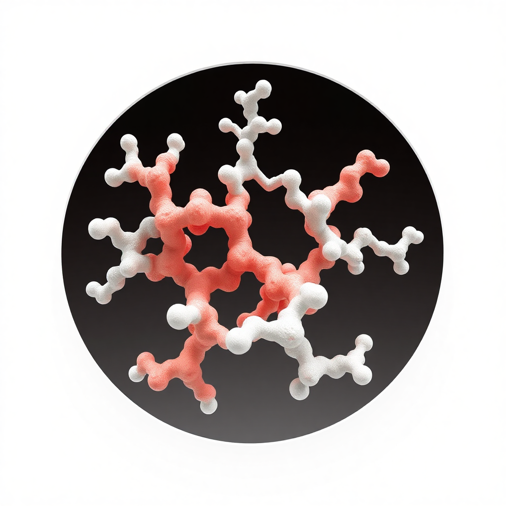
Enterokinase enzyme is a heterodimeric serine protease found in the duodenum of mammals. It has a molecular weight of 150 kDa, consisting of one heavy chain (115 kDa) and one light chain (35 kDa). It exhibits specific protein substrate hydrolysis within a pH range of 4.5-9.5 and a temperature range of 4-45°C.
As the purposeful peptides released by EK cleavage of fused proteins have an N-terminal amino acid sequence identical to the wild type, it can be used as a cutting reagent for fused proteins in protein expression systems. However, natural enterokinase enzyme sources are limited, and the cost of extraction and separation is high. In addition, natural extraction of enterokinase enzyme is easily contaminated by other proteases, resulting in degradation of the target product during the cutting of fused proteins. Genetic engineering methods can compensate for these shortcomings, and therefore, the use of genetic engineering methods to produce enterokinase enzyme is widely used. The two mainly commercialized enterokinase enzyme products are natural purified bovine enterokinase enzyme and genetically engineered recombinant bovine enterokinase enzyme.
Structural characteristics of enterokinase enzyme

Natural enterokinase enzyme consists of one structural subunit (heavy chain) and one catalytic subunit (light chain), which are linked by one intermolecular disulfide bond. The structural subunit is responsible for fixing the catalytic subunit on the brush border membrane in the small intestine and guiding it to move towards the intestinal lumen. The catalytic subunit can specifically recognize the Asp-Asp-Asp-Asp-Lys sequence and cleave it along the carboxyl end of the sequence, activating trypsinogen to trypsin enzyme, thus initiating cascades of enzyme activation.
The molecular weight of recombinant enterokinase is usually 26.3 kDa, with three glycosylation sites and a glycosylation molecular weight of approximately 43 kDa. In vitro experiments have shown that the enzyme has full enzymatic cleavage specificity and exhibits enhanced enzymatic cleavage activity towards gene-engineered fused protein substrates compared to natural enterokinase enzyme, making genetic engineering methods the preferred method of producing enterokinase enzyme. Recent attempts have focused on cloning and expressing a light chain of 235 amino acids.
The three-dimensional structure of the light chain is homologous to that of serine proteases such as trypsin. The enzyme activity is hardly affected when the Ile at the N-terminus of the light chain is mutated to Val. In addition, the heavy chain of EK strongly affects the recognition of enterokinase enzyme for macromolecular substrates, but has no effect on the recognition of synthetic fused proteins or small molecule substrates.
Enterokinase enzyme activates trypsinogen to produce trypsin enzyme in vivo
Since the light chain structure of enterokinase enzyme is conserved in humans, cattle, and pigs, the recognition sequence Asp-Asp-Asp-Asp-Lys also has strong conservation in vertebrates, and almost all sequenced trypsinogens have four consecutive N-terminal asparagine residues, which are rare in other natural proteins. The active center of enterokinase enzyme has a special cationic site, which allows the strongly negative Asp-Asp-Asp-Asp to bind to the site, making the substrate cleavage site sequence of bovine enterokinase enzyme highly specific. This feature makes EK a useful tool in the post-modification process of gene-engineered fused proteins. However, in actual enzymatic cleavage reactions, non-specific cleavage may also occur, which should be carefully addressed in gene engineering research.


